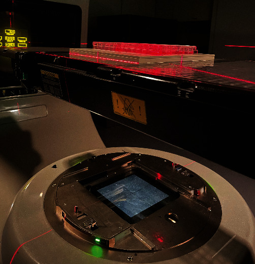
Figure 1.

Diagram of the X-ray radiation of HLECs. Plates seeded with HLECs were placed in a 30 × 30 cm2 field on 1.5 cm of equivalent tissue material, and the radiation (4 Gy of X-ray) was delivered using Varian linear accelerator (Clinac-21EX, USA).
