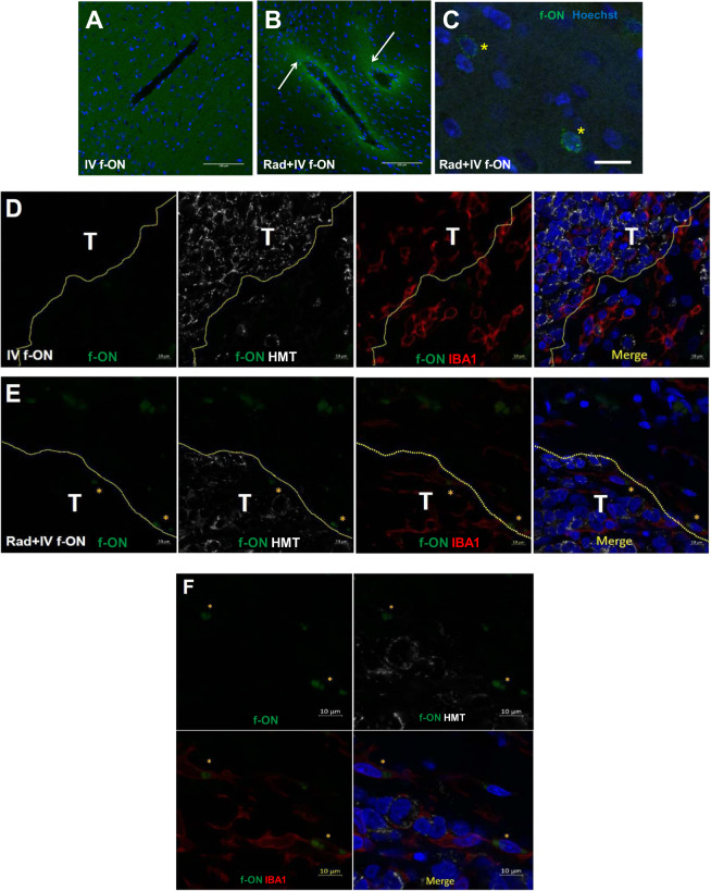
Fig. 2. Non-ablative radiation enhanced the delivery of f-ON into rat brain in vivo.
A–C Long Evan rats received IV f-ON (10.5 mg/kg; IV) either alone (A) or 1 day after 5 Gy whole-brain irradiation (B), (C). Brains were harvested 1 day after f-ON administration. Sections were counterstained with Hoechst nuclear stain and fluorescein localization was analyzed. C High magnification field showing punctate cellular uptake of f-ON. D–F Athymic nude rats with D283 tumor received f-ON alone (D) or 1 day after 2 Gy brain irradiation observed; representative micrographs under low magnification (E) or high magnification (F). Brains were harvested 1 day after f-ON administration and sections were stained for human mitochnondrial antigen (HMT) and ionized calcium-binding adapter molecule 1 (IBA1) as a marker of human tumor cells and microglia. Scale bar = 100 μm (A, B), 20 µm (C), or 10 µm (D, E, F), respectively. (T tumor and *representative f-ON positive cells).

