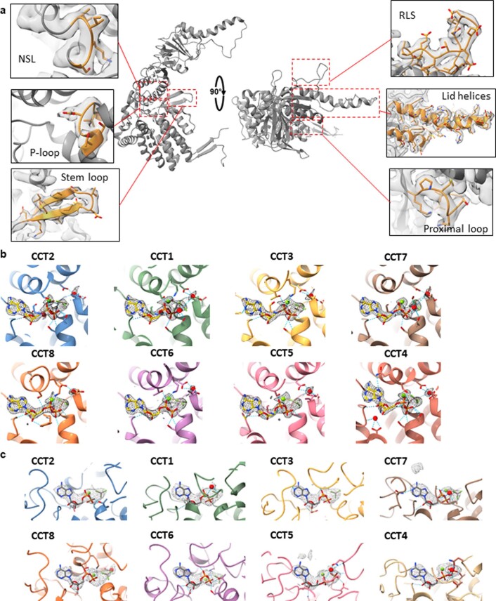
Extended Data Fig. 6. Conserved structural features of TRiC.
(a) Conserved structural motifs common to all CCT subunits (here represented by CCT1) including apical lid helices, proximal loop, release loop of substrate (RLS), P-loop, nucleotide-sensing loop (NSL), and stem loop. Insets: Motifs are coloured orange and density map is shown grey. (b,c) Snapshots of ligand density in the nucleotide binding site for each CCT subunit of closed-TRiC (b) and open-TRiC (c). Dashed lines represent hydrogen bonding. ADP molecules are shown in yellow with Mg2+ in green and water molecules are represented in red spheres.

