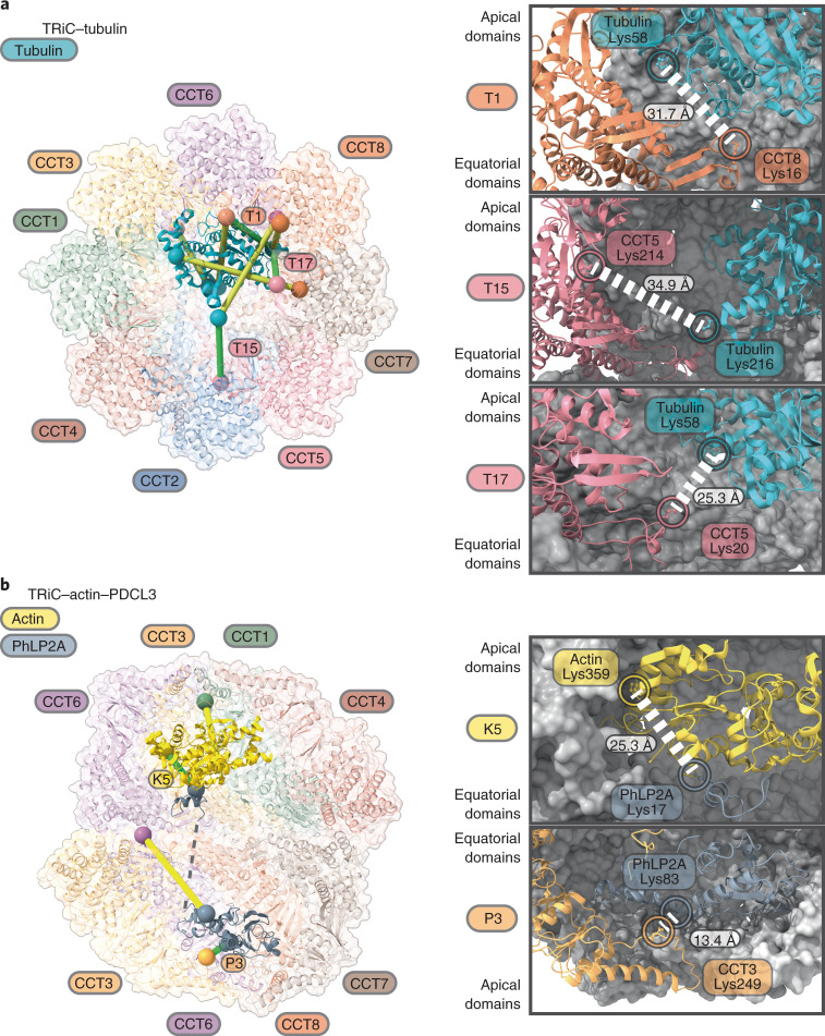
Fig. 5. Crosslinking mass spectrometry analysis of TRiC-substrate interactions within the folding cavity.
a,b, Crosslinks between TRiC and tubulin (a) and between TRiC and actin/PhLP2A (b) reported from literature are mapped onto the TRiC structure with bound proteins. Left, identified crosslinks compatible with our structural models are indicated in green lines; crosslinks that are not sterically compatible with our model are indicated in yellow lines and could represent other stages of the substrate folding cycle that are not captured in our structures. Spheres represent lysine residues that are crosslinked. Right, magnified views showing the atomic environment of 3 TRiC–tubulin, 1 actin-PhLP2A, and 1 TRiC-PhLP2A crosslinks that support our models.

