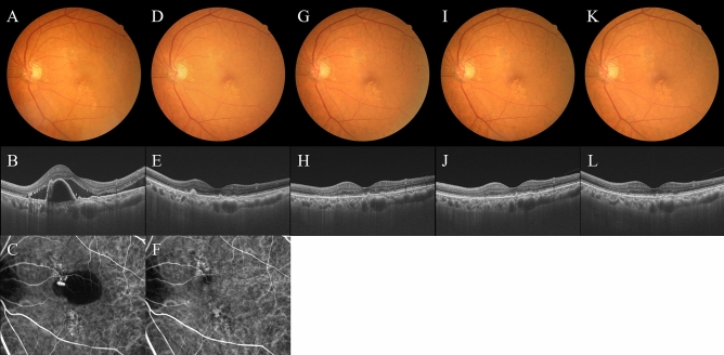
Figure 6.
Images of the left eye of a 76-year-old man with polypoidal choroidal vasculopathy. The results of loading phase treatment with brolucizumab for this eye were shown in our prior report8. At baseline, best-corrected visual acuity (BCVA) was 0.22 logarithm of the minimum angle of resolution (logMAR) units. (A) Color fundus photograph shows retinal pigment epithelium (RPE) degeneration and detachment accompanied by serous retinal detachment (SRD) at the macular area. (B) 12 mm B-mode optical coherence tomography (OCT) image through the fovea and polypoidal lesion shows sharply peaked RPE detachment due to the polypoidal lesion, which is accompanied by serous RPE detachment and SRD. Moreover, choroidal thickening associated with dilatation of outer choroidal vessels is seen in the area of the double layer sign reflecting a branching neovascular network. The foveal thickness and CCT are 388 μm and 360 μm, respectively. (C) Indocyanine green angiography (ICGA) shows polypoidal lesions in the RPE detachment and a branching neovascular network. At week 12, 4 weeks after the third injection of brolucizumab: BCVA of the left eye is -0.08 logMAR units. (D) Color fundus photograph shows RPE degeneration at the macular area. (E) 12 mm B-mode OCT image shows neither serous RPE detachment nor SRD. The foveal thickness and CCT are 148 μm and 303 μm, respectively. (F) ICGA shows no polypoidal lesions. At week 16: BCVA of the left eye is -0.08 logMAR units. (G, H) Color fundus photograph and 12 mm B-mode OCT image shows no recurrence of exudative changes. The foveal thickness and CCT are 165 μm and 313 μm, respectively. The fourth injection of brolucizumab was administered with an interval of 8 weeks. At week 28: BCVA of the left eye is -0.08 logMAR units. (I) (J) Color fundus photograph and 12 mm B-mode OCT image shows no recurrence of exudative changes. The foveal thickness and CCT are 146 μm and 313 μm, respectively. The fifth injection of brolucizumab was administered with an interval of 12 weeks. At week 44: BCVA of the left eye is − 0.08 logMAR units. (K, L) Color fundus photograph and 12 mm B-mode OCT image show no recurrence of exudative changes. The foveal thickness and CCT are 162 μm and 312 μm, respectively. The sixth injection of brolucizumab was administered with an interval of 16 weeks. The intended interval for the next injection of brolucizumab is also 16 weeks. No adverse events such as intraocular inflammation were observed during the 1-year treatment period.

