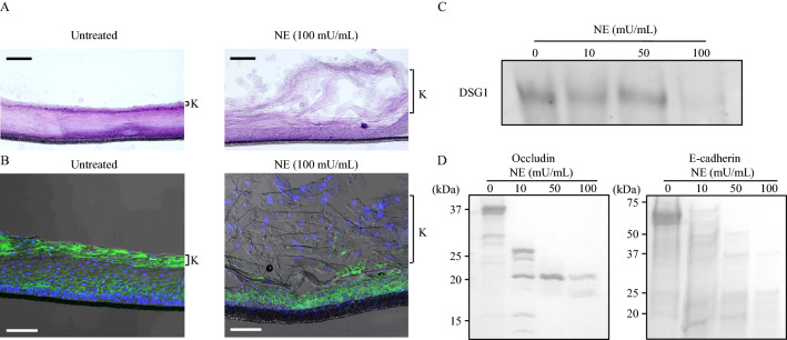
Figure 4.
Neutrophil elastase induces exfoliation of gingival epithelial keratinous layer via cleavage of cell adhesion molecule. (A, B) Three-dimensional human oral epithelial tissue models were exposed to 100 mU/mL NE followed by 12-h incubation. (A) Frozen sections were stained with hematoxylin and eosin. Representative images obtained by optical microscopy are shown. Scale bar: 100 μm. (B) Representative fluorescence microscopy images of the sections stained for DAPI (nucleus, blue) and DSG1 (green) are shown. Scale bar: 100 μm. K in Fig. 5A and B indicates keratinous layer. (C, D) Recombinant human DSG1, occludin, and E-cadherin were exposed to NE (10, 50, and 100 mU/mL) for 3 h and determined by western blotting (C) or Coomassie Brilliant Blue staining (D). DSG1 desmoglein 1, NE neutrophil elastase.

