Figure 3.

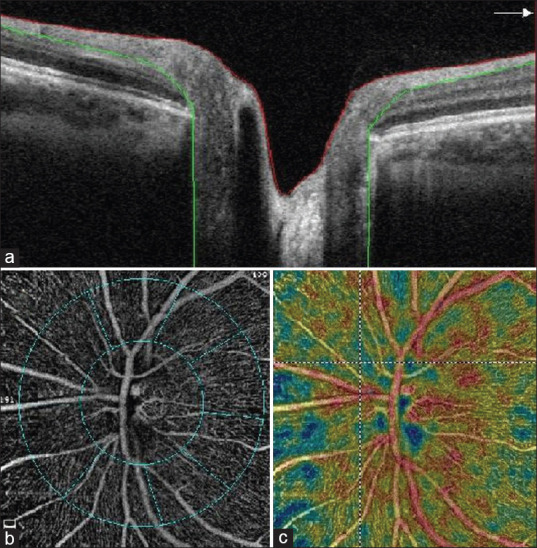
Peripapillary retinal nerve fiber layer was evaluated in the region extending from ILM (red line) to nerve fiber layer (NFL) (green line) (a), subdivisions of the peripapillary region (b), color image of the peripapillary region (c)

Peripapillary retinal nerve fiber layer was evaluated in the region extending from ILM (red line) to nerve fiber layer (NFL) (green line) (a), subdivisions of the peripapillary region (b), color image of the peripapillary region (c)