
Figure 2.
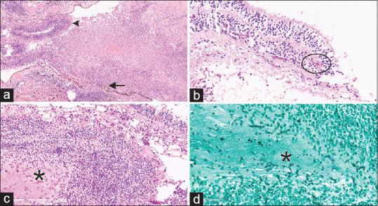
Photomicrograph of endogenous endophthalmitis. (a) Fragments of the choroid (arrow) and retina (arrowhead) densely infiltrated with inflammatory cells, extending into the vitreous cavity (H and E, ×200). (b) Fragments of fungal hyphae (within the circle) noted in the retina (H and E, ×300). (c and d) Suppurative inflammation of vitreous (asterix) with fungal hyphae, which are hyaline, thin, septate, and branched (PAS left × 300 and GMS right, ×400)