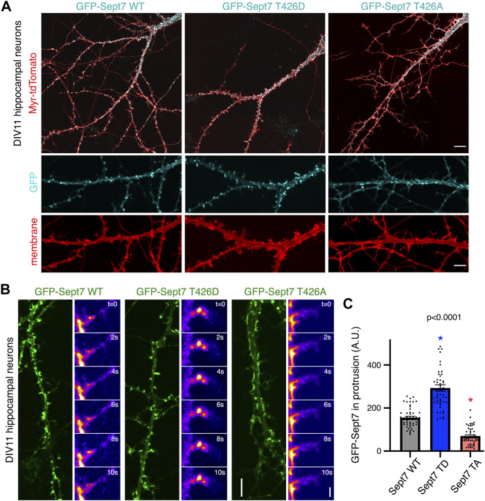
FIGURE 2.
Phosphorylation of Sept7 alters its localization and dynamics. (A) Hippocampal neurons were transfected with GFP-tagged Sept7-WT, phosphomimetic Sept7-T426D, or phosphomutant Sept7-T426A along with membrane marker myristoylated-tdTomato at DIV9 and then fixed at DIV11. Phosphomimetic GFP-Sept7 (T426D) expression leads to early maturation of dendritic spines compared to Sept7-WT, while phosphomutant Sept7-T426A expressing neurons exhibited only filopodial protrusions. (B) Montage of time lapse images showing DIV11 hippocampal neurons transfected at DIV9 with GFP-tagged wild-type Sept7-WT, phosphomimetic Sept7-T426D, or phosphomutant Sept7-T426A. Phosphomimetic Sept7-T426D is stably localized within the dendritic spine head, phosphomutant Sept7-T426A localizes to the base of the dendritic spine, whereas the wild-type Sept7-WT shows dynamic changes in its localization within protrusions. Scale bar represents 5 µm (green) and 1 µm (montage), respectively. (C) Quantification of GFP intensity in protrusions of DIV11 neurons transfected with GFP-tagged wild-type Sept7-WT, phosphomimetic Sept7-T426D, and phosphomutant Sept7-T426A. Values represent mean intensity, error bars represent SEM and n = 50 protrusions from 10 neurons in each condition. Dunnett’s multiple comparison test, p < 0.0001.

