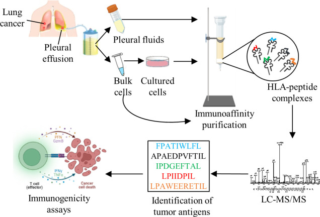
Figure 1.
Experimental design. Pleural effusions were collected from cancer and non-cancer patients, cells were separated and cultures were grown, when possible. HLA-peptide complexes were immunoaffinity-purified, peptides were separeted and analyzed by mass spectrometer. Peptides were considered HLA ligands after filtration of known contaminants, and if they were 8–14 amino-acids-long and matched the HLA allele consensus motifs of each patient, based on their tissue typing and online analysis with the NetMHCpan V.4.1 server http://www.cbs.dtu.dk/services/NetMHCpan/. Tumor antigens were identified from the peptides lists and candidates were tested for an immunogenic response. The figure was created using BioRender (https://biorender.com/). HLA, human leucocyte antigen; LC-MS/MS, liquid chromatography and tandem mass spectrometry.

