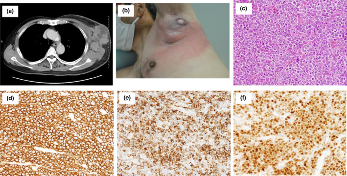
Figure 1.

Clinical and histopathological characteristics of case 1. (a) CT image revealed enlarged mass in the left axilla. (b) A clinical image of the case 1 at the biopsy was presented. (c) An image of Haematoxylin and Eosin staining was shown (×200). (d–f) Images of immunohistochemical staining for CD20 (d × 100), BCL‐2 (e × 100) and MUM1/IRF4 (f × 100) were shown. [Colour figure can be viewed at wileyonlinelibrary.com]
