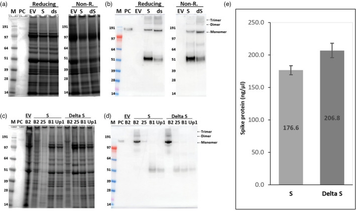
Figure 7.

Expression and purification of the S protein of SARS‐CoV‐2 Wuhan strain and Delta variant. SDS–PAGE and Western blot against S protein using protein extracts (a, b) and fractions from sucrose cushions (c, d) of leaves infiltrated with S and Delta S (dS). The S protein concentration in fraction# 6 samples from iodixanol gradients of the S and dS samples was measured by ELISA (e). Error bars indicate the standard deviation of three different wells. Lane M: Protein size marker; Lane PC: 100 ng (for Western blot) or 300 ng (for staining) of SARS‐CoV‐2 spike protein from CHO cells as positive control; reducing: samples boiled in the presence of β‐mercaptoethanol; non‐R: samples boiled in the absence of β‐mercaptoethanol; EV: empty vector controls processed using the same conditions as for S and dS; Lane B2: interface between 25 and 70% sucrose layers plus the 70% layer; Lane 25: 25% sucrose fraction; Lane B1: interface between the supernatant and 25% sucrose fraction; Lane up1: supernatant above the sucrose cushions.
