FIGURE 2.

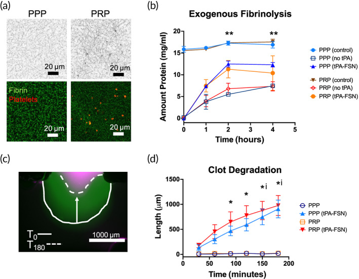
Clot degradation with tissue‐type plasminogen activator‐fibrin‐specific nanogels (tPA‐FSNs) in platelet‐rich and platelet‐poor plasma. (a) Confocal microscopy of platelet‐rich plasma (PRP) and platelet‐poor plasma (PPP) illustrating enhanced fibrin clot structure in platelet‐rich environments compared to platelet‐poor environments. Fibrin fibers are depicted in black (top) and green (bottom), and platelets are shown in red (bottom). (b) Exogenous fibrinolysis assays show enhanced degradation with overlayed tPA‐FSNs compared to no tPA. **p < 0.01 for PPP (no tPA) compared to PPP (tPA‐FSNs). (c) Clot degradation studied in T‐junction fluidic devices is shown as overlayed images at T0 (after binding of tPA‐FSNs for 20 min at a wall shear rate of 1 s−1) and T180 at the stationary clot boundary. (d) Quantification of clot degradation of PPP and PRP stationary clots with and without tPA‐FSNs in flow solutions for 20 min prior to degradation study (n = 3 clots per condition). Mean ± SD is shown. * = p < 0.05 between PPP conditions. i = p < 0.05 between PRP conditions. Data were analyzed via a two‐way analysis of variance with a Tukey's post hoc test using a 95% confidence interval
