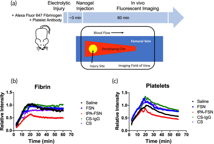
FIGURE 3.

Clot dynamics in vivo with tissue‐type plasminogen activator (tPA)‐loaded nanogels. (a) Schematic of electrolytic injury induced in mice followed by nanogel injections (~3 min after injury) and intravital fluorescent imaging continuously monitoring fibrin/fibrinogen and platelet recruitment at the injury site up to 60 min post‐injury. Treatment groups include saline, fibrin‐specific nanogels (FSNs), tPA‐FSNs, CS nanogels, and control sheep immunoglobulin G (CS‐IgG) nanogels (n = 4 animals/group). (b) Intravital microscopy quantification demonstrates accumulation of fibrin/fibrinogen at the injury site with less fibrin incorporation upon tPA‐FSN treatment. (c) Intravital microscopy quantification also demonstrates accumulation of platelets at the injury site and similarly shows reduced platelet incorporation with tPA‐FSNs. Data presented are mean fluorescent intensity plots over time (frame rate = 0.167 min)
