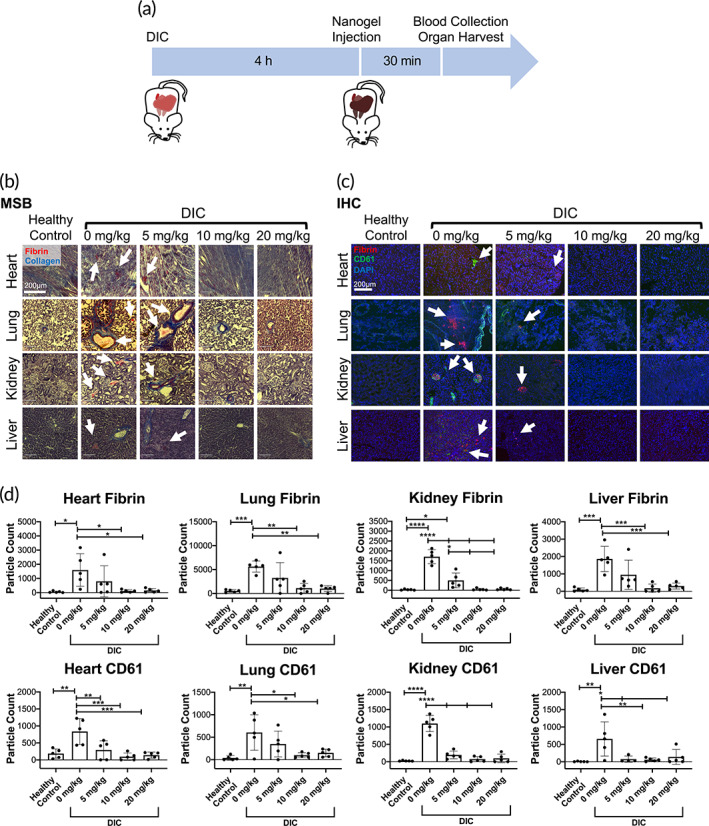
FIGURE 4.

Dose‐response of tissue‐type plasminogen activator‐fibrin‐specific nanogel (tPA‐FSN) treatment and thrombi presentation in a disseminated intravascular coagulation (DIC) rodent model. (a) Schematic of the DIC animal model used including treatment and terminal endpoints. (b) Martius scarlet blue (MSB) stained tissue sections of heart, lung, kidney, and liver from control animals and DIC animals treated with 0, 5, 10, and 20 mg/kg tPA‐FSNs (fibrin = red, collagen = blue). (c) Immunohistochemistry (IHC) tissue sections of heart, lung, kidney, and liver from control animals and DIC animals treated with 0, 5, 10, and 20 mg/kg tPA‐FSNs (fibrin = red, CD61 = green, DAPI = blue). (d) Quantification of IHC fibrin and platelet (CD61) presentation in tissue sections via ImageJ particle count analysis. For each organ, at least three quantified tissue sections were measured and averaged per animal. The mean of five animals per group ± SD is shown. Data were analyzed via a one‐way analysis of variance with a Tukey's post hoc test using a 95% confidence interval. *p < 0.05, **p < 0.01, ***p < 0.001, ****p < 0.0001
