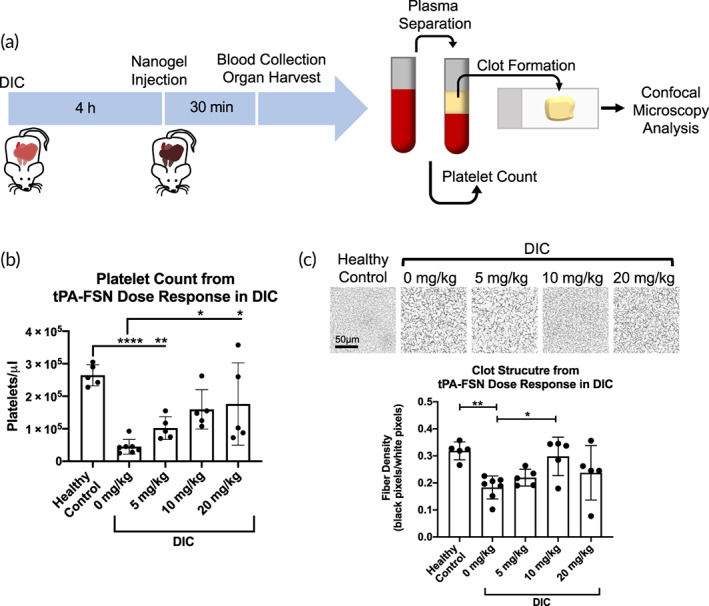
FIGURE 5.

Platelet count dose‐response and clot structure measured after tissue‐type plasminogen activator‐fibrin‐specific nanogel (tPA‐FSN) treatment in a disseminated intravascular coagulation (DIC) rodent model. (a) Schematic of the DIC animal model used in addition to the treatment timing and terminal endpoints. Terminal blood collection was followed by platelet counts and plasma separation to form ex vivo clots and examine clot structure at the study endpoint. (b) Manual platelet counts were conducted for control and DIC animals treated with 0, 5, 10, and 20 mg/kg tPA‐FSNs. (c) Confocal microscopy images were taken, and fiber density quantification was conducted of clots polymerized from isolated platelet‐poor plasma from control animals and DIC animals treated with 0, 5, 10, and 20 mg/kg tPA‐FSNs. Clots from each animal were polymerized and three images per clot were quantified for fiber density and averaged. n = at least 5 animals/group. Mean ± SD is shown. Data were analyzed via a one‐way analysis of variance with a Tukey's post hoc test using a 95% confidence interval. *p < 0.05, **p < 0.01, ****p < 0.0001
