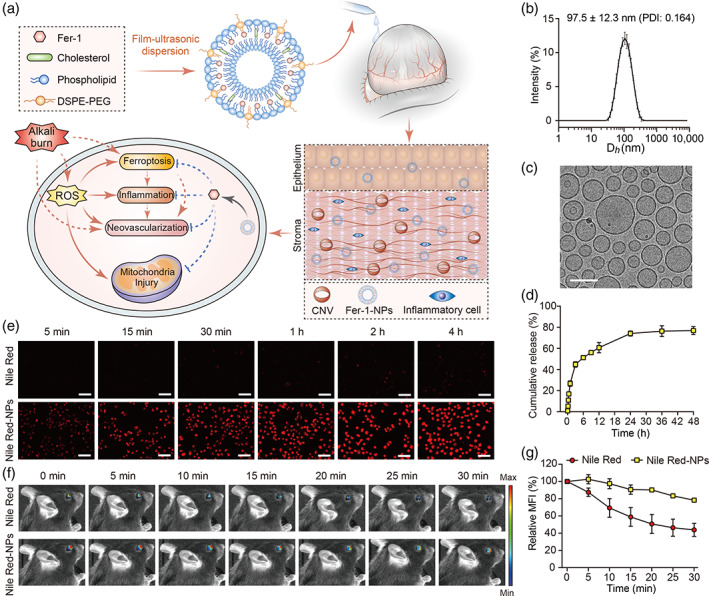
FIGURE 3.

Self‐assembly and characterization of Fer‐1‐loaded liposomes and their predicted effects on corneal cells after alkali burn injury. (a) Liposomes were prepared using a thin‐film hydration method with Fer‐1 encapsulated into the hydrophobic layer. When administrated to the alkali‐burned cornea by eye drops, nanocarriers‐encapsulated Fer‐1 exhibit enhanced drug bioavailability than free Fer‐1. After the internalization of Fer‐1‐NPs, the drugs are released inside the cell, where they exert their inhibitory effects of ferroptosis, inflammation, and neovascularization. (b) The dynamic light scattering‐determined hydrodynamic diameter of the Fer‐1‐NPs (n = 3). (c) The cryo‐transmission electron microscope image of the Fer‐1‐NPs; scale bar, 100 nm. (d) In vitro release profiles of the Fer‐1‐NPs (n = 3). (e) Cellular uptake of free Nile Red and Nile Red‐NPs by human corneal epithelial cells. Scale bar, 50 μm. Representative fluorescence images of mice eyes (f) and quantification (n = 3) of the fluorescence signal (g) at different time points after topical administration of free Nile Red or Nile Red‐NPs. Results were presented as the mean ± SD. CNV, corneal neovascularization; DSPE‐PEG, 1,2‐distearoyl‐sn‐glycero‐3‐phosphoethanolamine‐N‐[methoxy (polyethylene glycol)‐2000]; Fer‐1‐NPs, ferrostatin‐1‐loaded liposomes; PDI, polydispersity index; ROS, reactive oxygen species
