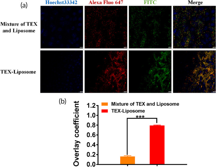
FIGURE 3.

Verification of TEX‐Liposome generation. (a) CLSM images of cellular uptake of untreated mixture and TEX‐Liposome nanoparticles after incubation with NIH3T3 cells for 4 h. Green for FITC labeled Liposome which was prepared by DSPE‐PEG‐FITC phospholipid. Red for Alexa Fluor 647‐anti‐CD9 (TEX specific membrane protein) antibody labeled TEX membrane. The nucleus was indicated using Hoechst 33342 (Blue). Scale bars = 10 μm. (b) The overlay coefficient analysis according to (a) using Image J. ***p < 0.001. TEX, tumor‐derived exosome
