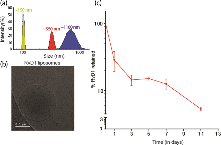
FIGURE 1.

Characterization and release profile of Lipo‐RvD1. (a) The size distribution of liposomes used in this study as measured using dynamic light scattering. (b) Cryo‐TEM micrographs of Lipo‐RvD1. (c) Quantification of in vitro release of RvD1 from Lipo‐RvD1 when incubated at 37°C at pH 7.4; n = 3. Cryo‐TEM, cryogenic transmission electron microscope
