FIGURE 1.

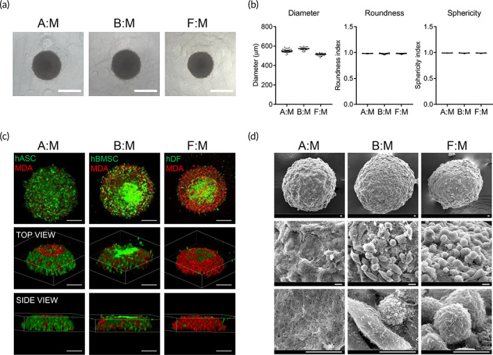
Formation and morphological characterization of 3D multicellular tumor spheroids. (a) Representative phase‐contrast images of tumor spheroids at 2 days after seeding with densities of 10,000 cells/well. Scale bar, 500 μm. (b) Shape properties of tumor spheroids. n = 40 per group. (c) Distribution of cancer cells and stromal cells in the tumor spheroids. Stromal cells (hASCs, hBMSCs, or hDFs) and breast cancer cells (MDA‐MB‐231) were stained with green and red fluorescent dyes, respectively. Scale bars, 200 μm. (d) SEM images of tumor spheroids, scale bars, 10 μm. A:M, B:M, or F:M; multicellular tumor spheroids generated using hASC, hBMSC, or hDF along with MDA‐MB‐231, respectively. 3D, three dimension; hASCs, human adipose‐derived stromal cells; hBMSCs, human bone marrow stromal cells; hDFs, human dermal fibroblasts; SEM, standard error of the mean
