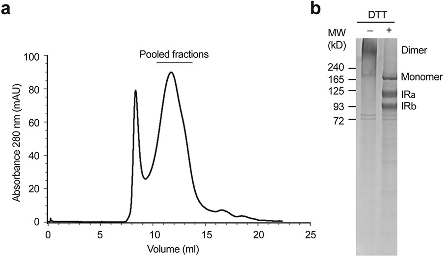
Extended Data Fig. 2. Purification of the full-length mouse insulin receptor (IR).
a. A representative size-exclusion chromatography of IR.
b. The peak fractions were combined and visualized on SDS-PAGE by Coomassie staining, in the absence or presence of dithiothreitol (DTT). Most of IR was processed into α-chain (IRα) and β-chain (IRβ). This experiment was repeated for 10 times independently with similar results.

