
Figure 4: Satellite-derived ncRNAs organize HP1 at inter-chromosomal hubs.
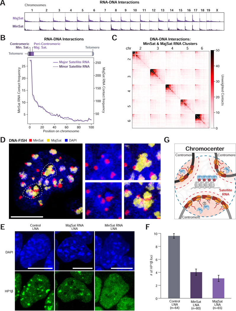
(A) Unweighted RNA-DNA contact frequencies of major and minor satellite-derived ncRNAs across the genome or (B) aggregated across all chromosomes. (C) Unweighted DNA-DNA contacts for chromosomes 2 – 6 within clusters containing a satellite-derived RNA. (D) DNA FISH of major (yellow) and minor (red) satellite DNA in the nucleus (DAPI, blue). Dashed lines demarcate the two DAPI-dense structures shown as zoom-ins on the right. Scalebar is 10μm. (E) HP1β IF following LNA-mediated knockdown of major (MajSat) and minor (MinSat) satellite-derived RNAs. Scalebar is 10μm. (F) Quantification of the mean number of HP1β foci per cell following LNA knockdown. n=number of cells analyzed, error bars represent standard error. (G) Schematic of Chromocenter Hub. Satellite RNAs are spatially concentrated (red gradient) near centromeric DNA. Individual centromeres assemble into a heterochromatic chromocenter structure highly enriched with HP1 protein. See also Figure S4.