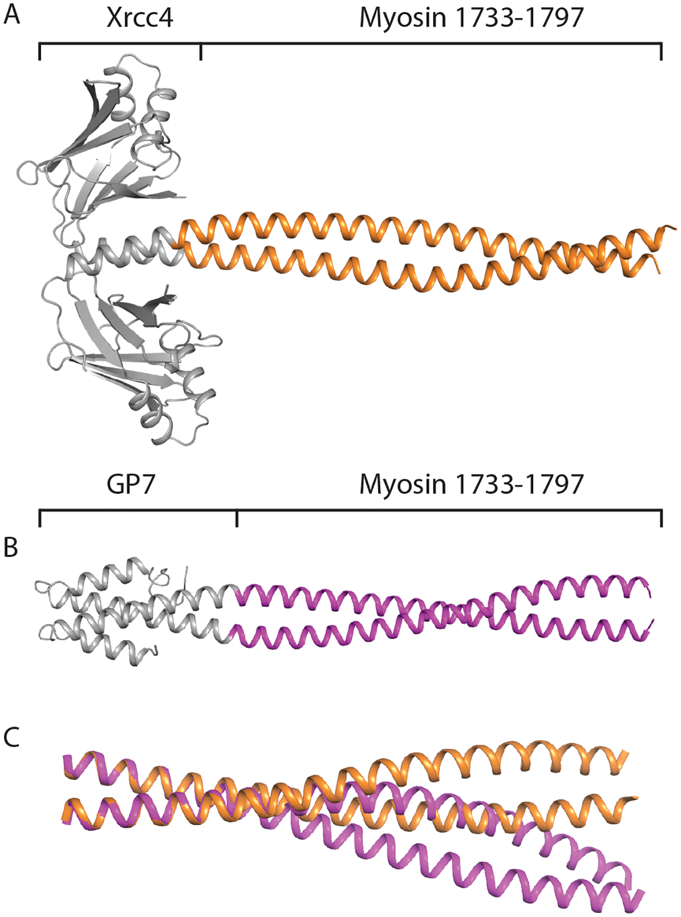
Figure 4. Structures and comparison of the dimeric fusion proteins covering.

Asp1733 - Leu1797. (A) Structure of the coiled-coil domain of myosin rod from amino acids 1733–1797 fused to Xrcc4 fusion domain. (B) Structure of myosin rod from amino acids 1733–1797 fused to Gp7 fusion domain. (C) Aligned structure of the coiled-coil domains of the Xrcc4-1733-1797 structure and Gp7-1733-1797 structure. The two conformations of myosin were aligned from amino acids 1733–1746, and the deviation in coiled-coil pitch is clearly visible between the two conformations.
