
Fig. 4.
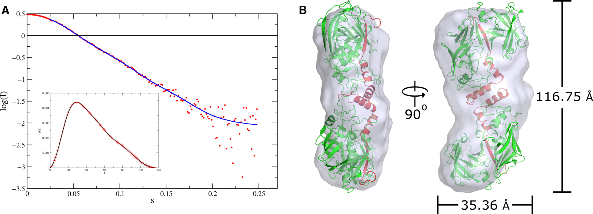
Dimeric nature of pro-tPMs in solution as revealed by SAXS experiment. (A) The scattering curve of pro-tPMI. The inset shows the distance distribution function. (B) Superimposed S-shaped dimeric model of pro-tPMI in the SAXS envelope (gray). The radius of gyration and maximum dimension of the envelope obtained from SAXS data are also shown. The model of dimeric pro-tPMI was fitted in the envelope using DAMMIN [54]. The protein structural representations have been generated with the PyMOL molecular graphics system (Schrödinger LLC, New York, NY, USA; version 2.3.2).