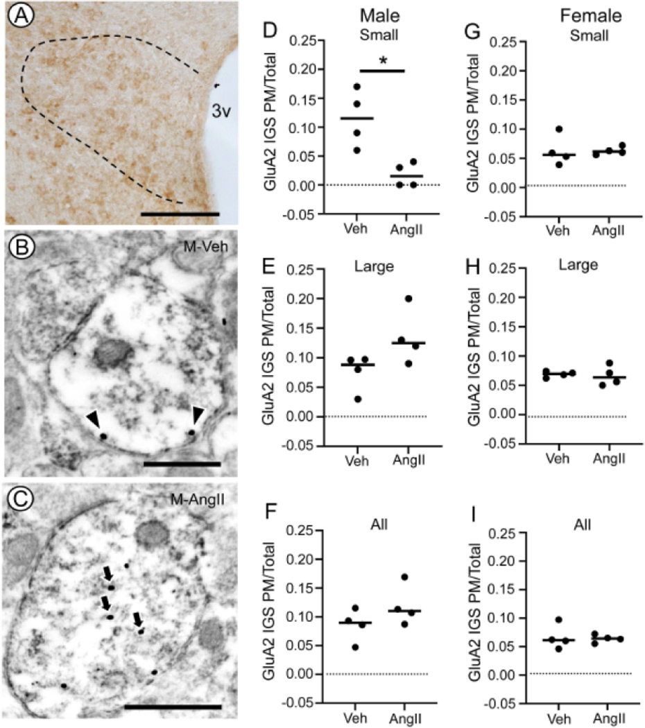
Fig. 5. Plasma membrane GluA2 IGS in TNFR1-labeled dendritic profiles of PVN neurons after AngII infusion in male and female mice.
Light micrograph illustrating GluA2 labeling in the dorsal hypothalamus of a vehicle-infused male mouse (A). The PVN is indicated by the bounded region. Electron micrographs illustrating GluA2 immunogold-silver (IGS) labeling in the cytoplasm (arrows) and on the plasma membrane (arrowheads) of immunoperoxidase TNFR1 labeled dendritic profiles of PVN neurons from male mice infused with vehicle (M-Veh, B) or AngII (M-AngII; C). Male mice infused with AngII showed a lower partitioning ratio for plasma membrane GluA2 IGS in small-size dendritic profiles compared to Veh-infused mice (D). There were no differences in the plasma membrane partitioning ratio of GluA2 IGS in large (E) profiles or profiles collapses across all sizes (F). In female mice, there were no differences in the plasma membrane partitioning ratio of GluA2 in small (G) or large (H) dendritic profiles. There also was no difference in plasma membrane GluA2 in dendrites collapsed across size (I). 3v: Third ventricle; M-AngII: Male AngII treatment; M-Veh: Male Veh treatment; PM/Total: Plasma membrane labeling/Total labeling. D: * p< 0.05 GluA2 IGS PM/Total Veh versus AngII in small dendrites in males. Lines in D-I are group medians overlain with data points representing means for each animal. Scale bars: A: 500 μm; B and C: 500 nm

