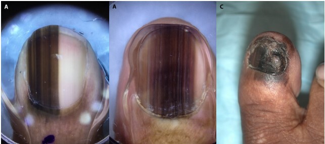
Figure 2.

Nail melanoma cases. (A,B) The dermoscopic images of two melanoma-in-situ cases that demonstrate micro-Hutchinson sign and brown to black parallel lines on the nail plate with irregular spacing, thickness, and disruption of parallelism. (C) The clinical image of an invasive melanoma case that demonstrates and easily visible Hutchinson sign, diffuse pigmentation of the whole nail plate, and nail dystrophy.
