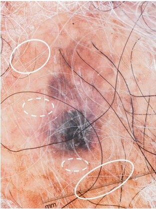
Figure 3.

Loss of normal pigmented network surrounding the squamous cell carcinoma lesion. Note the pigmented network seen in the normal skin surrounding the pigmented squamous cell carcinoma (solid ovals) and the loss of network just adjacent to the lesion (dashed ovals). This was observed in 4 cases (57.1%).
