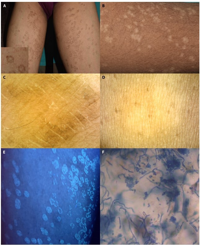
Figure 1.

(A, B) Multiple discrete scaly annular patches with peripheral rim of hyperpigmentation and central hypopigmentation over bilateral thighs. (C) Dermoscopy (captured with polarized dermoscopy, magnification × 10) revealed brown scales at the periphery of the lesion with white fine scales at the center within the skin creases. (D) On wiping the lesion with an alcohol swab, dermoscopic analysis showed complete disappearance of brown scales in the periphery with pigment dilution in the center. (E) Yellowish green fluorescence seen at Woods lamp examination (F) KOH mount shows fungal spores and hyphae (spaghetti and meatball appearance).
