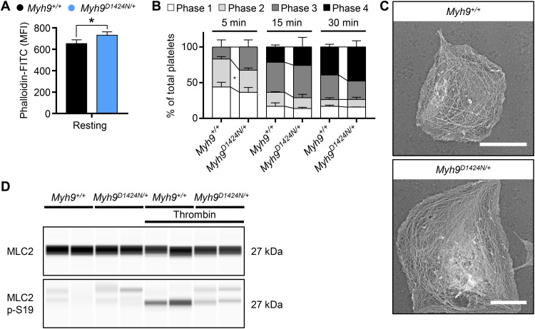
Fig. 2. Phosphorylation of MLC2 is decreased in Myh9D1424N/+ platelets.
(A) F-actin content of resting platelets was measured by flow cytometry after incubation with phalloidin-FITC (n = 3 to 7). The MFI is shown. (B) Statistical analysis of the different spreading phases (phase 1, resting platelets; phase 2, platelets forming filopodia; phase 3, platelets forming lamellipodia and filopodia; and phase 4, fully spread platelets) of fixed Myh9+/+ and Myh9D1424N/+ platelets on fibrinogen at different time points expressed as mean ± SD (n = 2). (C) Representative PREM (platinum replica electron microscopy) images of the cytoskeleton ultrastructure of platelets spread on fibrinogen in the presence of thrombin (scale bars, 2 μm). (D) Expression of MLC2 and phosphorylated MLC2p-S19 in resting and thrombin-activated (0.05 U/ml, 1 min) platelets was determined by using an automated quantitative capillary-based immunoassay platform, Jess (ProteinSimple). Representative immunoblot of three independent experiments is shown (n = 2). Statistics: multiple comparison using Holm-Sidak (*0.05 > P ≥ 0.01).

