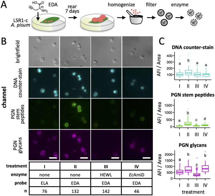
Fig 4. Labeling of A. pisum LSR1-c Buchnera cells with EDA by host feeding.
A) Aphids were reared on plant leaves immobilized in agar containing ethynyl-d-Ala (EDA) or the control l enantiomer from birth until the 4th instar larval stage, after which Buchnera cells were purified from aphid homogenate. B) Cells were fixed and stained with azide-linked Alexa Fluor 488 to label PGN stem peptides (PGN stem peptides), wheat germ agglutinin CF-640R conjugate to label the PGN glycan backbone (PGN glycans), and the counter-stain DAPI to label DNA (DNA counter-stain). Hen eggwhite lysozyme (HEWL; PGN glycan-targeting) and E. coli AmiD (PGN stem peptide-targeting) were used to demonstrate incorporation of EDA within Buchnera PGN stem peptides. Images were falsely colored to aid visualization. The white scale bar represents 5 μm. C) For each combination of enzyme and probe, the average fluorescence intensity (AFI) per area was measured for each of n Buchnera cells using ImageJ software (S9 Table). Significant differences between means are indicated by compact letter displays above each boxplot—means with no letters in common are significantly different by the Dunn’s Multiple Comparison test (p-value < 0.01 following adjustment for false discovery).

