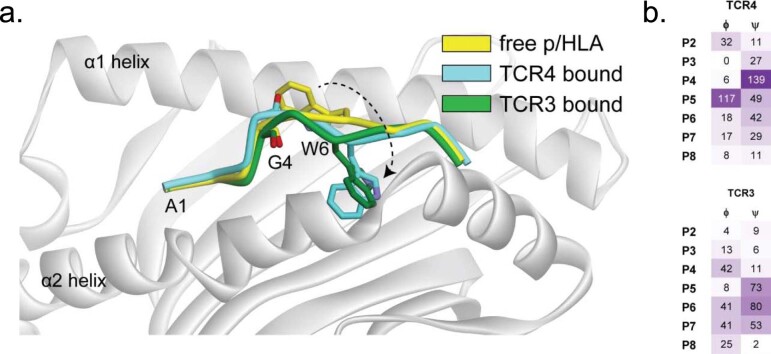
Extended Data Fig. 4. Mutant peptide conformational changes following TCR3 or TCR4 binding.
(a) Visualization of the conformations of the pMut backbone in the unbound state and upon binding of TCR3 and TCR4, emphasizing changes that occur in the positions of the P6 Trp side chain and the P4 Gly backbone carbonyl. (b) Table summarizing changes (in degrees) in the backbone dihedral angles (ϕ) and (ψ) of the pMut peptide following binding of TCR4 (top) or TCR3 (bottom). For binding of TCR4, the peptide conformational change is primarily driven by a change in the P4 ψ and P5 ϕ, whereas for TCR3 the change is driven by smaller dihedral changes spanning P4 to P7.

