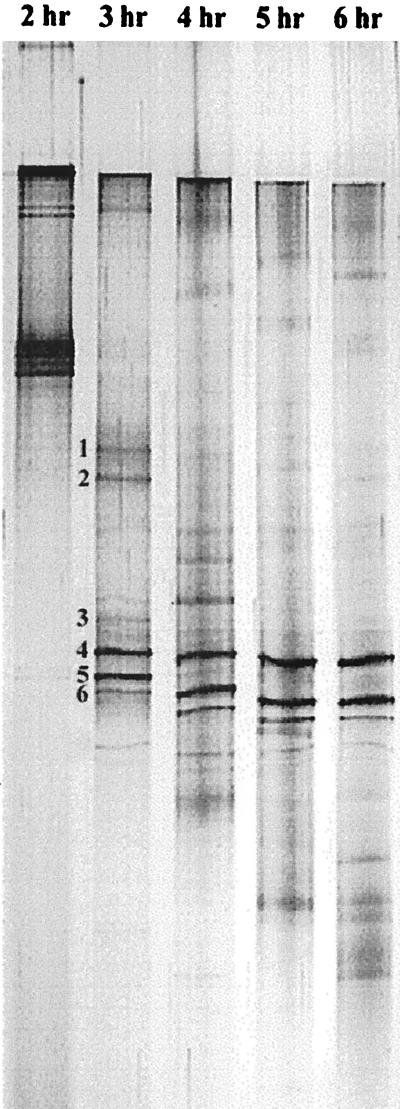
FIG. 3.
Silver-stained DGGE pattern of PCR-amplified 16S rDNA from the total DNA extracted from the deteriorated EBPR reactor. Electrophoresis was done for 2 to 6 h with a 1-h interval to optimize the running time. Up to 11 bands were clearly observed in the original gel. The six most dominant bands, which were further isolated and sequenced, are indicated next to the 3-h lane.

