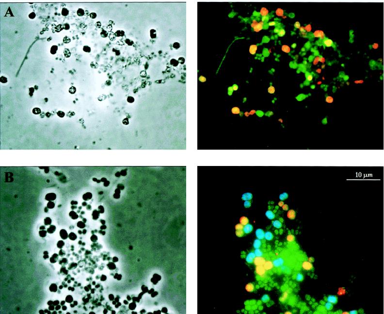
FIG. 5.
In situ hybridization of activated sludge from the deteriorated EBPR reactor. The left side shows phase-contrast images, and the right side shows epifluorescence micrographs of the corresponding areas. (A) In situ hybridization with probes specific for the domain Bacteria (green) and the gamma subclass of the Proteobacteria (red). (B) In situ hybridization with probes specific for the domain Bacteria (green) and for the novel subgroup of the gamma subclass of the Proteobacteria; the probe specific for the bacteria from band 4 is shown in blue, and the probe specific for the bacteria from bands 3, 5, and 6 is shown in yellow. The scale bar applies to all of the images.

