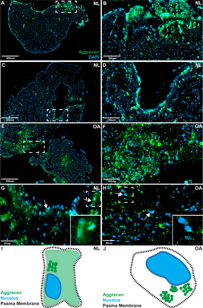
Fig. 4. Aggrecan staining in human synovium.
Normal (n = 8) and OA (n = 8) synovial biopsy samples were stained with aggrecan and DAPI. In normal (NL) synovium, aggrecan staining was predominantly observed within the intimal layer, but was also observed infrequently within the sub-intima (A–D two individuals shown as representative examples). In OA synovium, aggrecan staining was observed equally between the intima and sub-intima (E, F). The subcellular localization of aggrecan was also different between normal vs. OA samples. In normal samples aggrecan staining was diffuse throughout the cells with areas of increased staining adjacent to the nucleus (G arrows), while in OA samples a punctate pattern of staining was observed with increased staining adjacent to the nucleus (H arrows). Diagrammatic representations of aggrecan staining in normal (I) and OA (J) are also provided.

