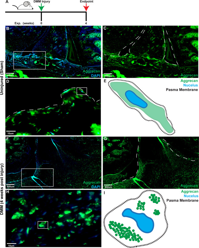
Fig. 5. Aggrecan staining in rat synovium.
Normal (n = 5) and DMM induced OA (n = 5) rat joints (A) were stained with aggrecan and DAPI. In normal synovium aggrecan staining was observed within the intimal and the sub-intimal (B, C) layers and was diffuse throughout the cell (D diagrammatic representation E). In OA synovium, aggrecan staining was throughout the intima and sub-intima (F, G). In OA synovium a punctate pattern of staining was observed in the synovium (H diagrammatic representation I).

