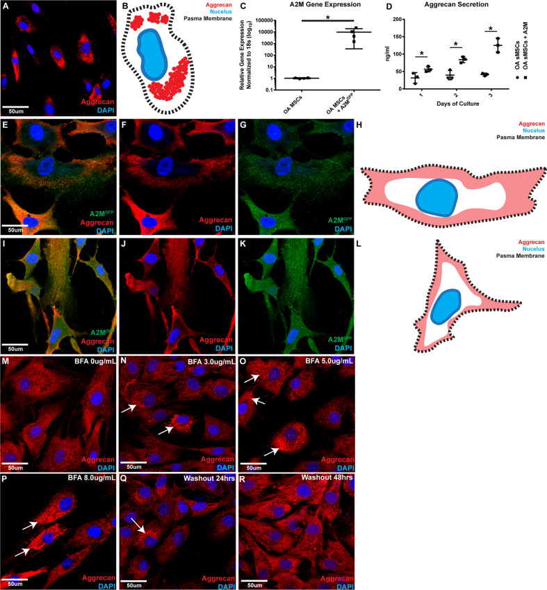
Fig. 7. Overexpression of A2M alters aggrecan staining pattern and increase secretion in OA synovial MPCs.
Un-transfected OA synovial MPCs demonstrating intense, punctate, perinuclear aggrecan staining pattern adjacent to the nucleus as observed previously (A diagrammatic representation B). OA MPCs also demonstrated low expression levels of A2M, and this could be rescued through A2M overexpression (C). In OA MPCs, A2M overexpression increased the secretion of aggrecan during all time-points examined when compared to the un-transfected parental cell lines (D). With A2M overexpression in OA MPCs (two patient cell lines shown as representative examples: E, I), aggrecan staining was no longer observed adjacent to the nucleus (F, J diagrammatic representation H, L). Instead A2M expression (GFP) was observed throughout the cytoplasm of the transfected cells (G, K). BFA treated of A2M transfected MPCs results in a redistribution of aggrecan within the cells directly related to the BFA concentration (M–P). This phenotype can be reversed by removing the BFA (Q, R). Scale bars equal 50 µm. *p < 0.05.

