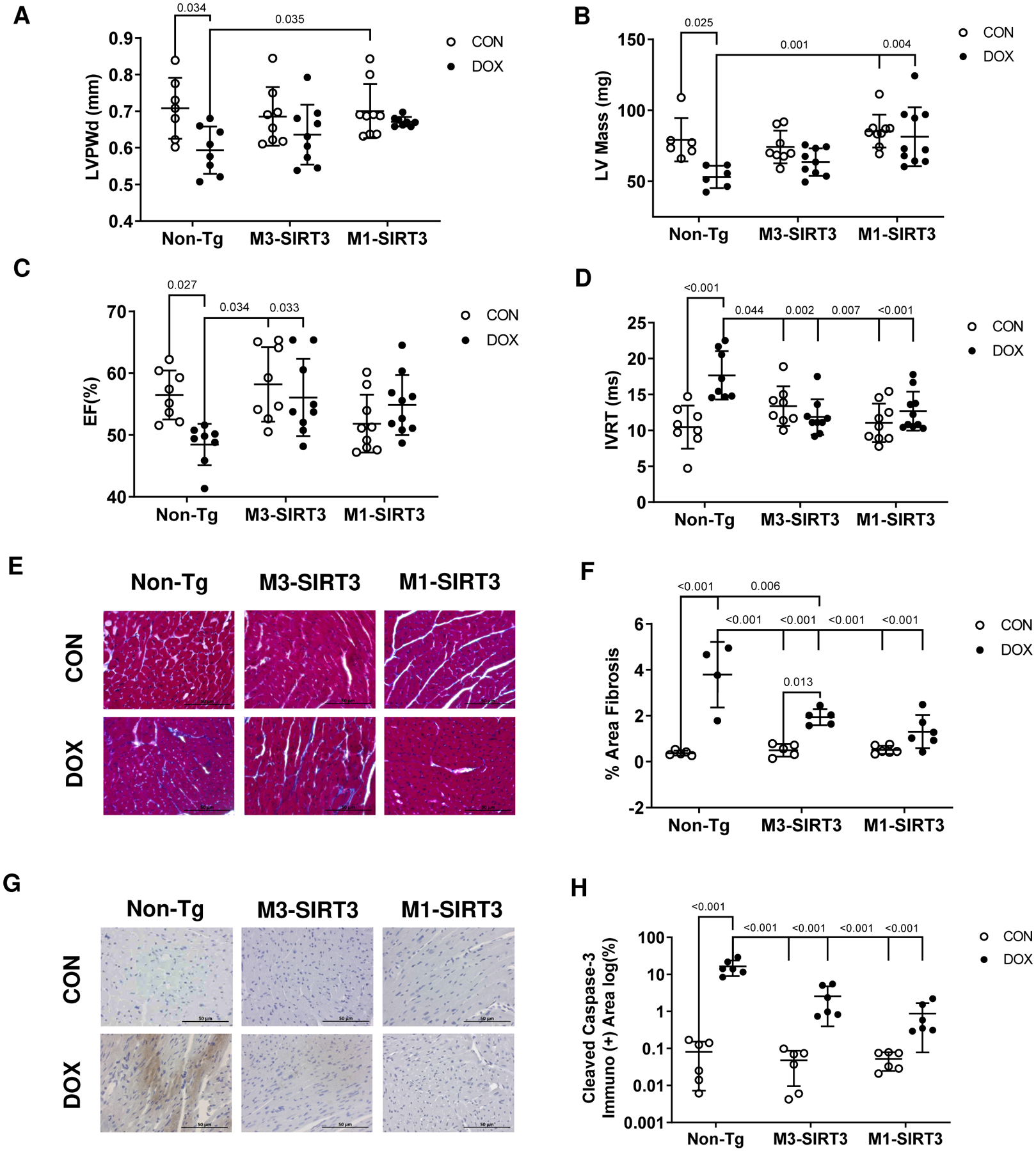
Figure 2. M1-SIR3 Transgenic Mice are Resistant to DOX Induced Cardiac Remodelling and Dysfunction.

M3-SIRT3, M1-SIRT3 and Non-Tg controls treated with 8.0mg/kg of DOX or saline once a week for four weeks. (A) Left ventricular posterior wall thickness during diastole. (B) Left ventricular mass. (C) Ejection fraction. (D) Isovolumetric relaxation time (n=8–10) (E) Representative images of trichrome staining. (F) Area fibrosis quantification of trichrome staining (n=4–6) (G) Representative images of cleaved caspase-3 staining. (H) Area quantification of immuno-positive cleaved caspase-3 immunohistochemistry (n=6). Scale bars = 50μm. Sample size refers to biological replicates. Female mice. Values are mean ± SD. Statistics are two-way ANOVA with Tukey’s post-hoc test.
