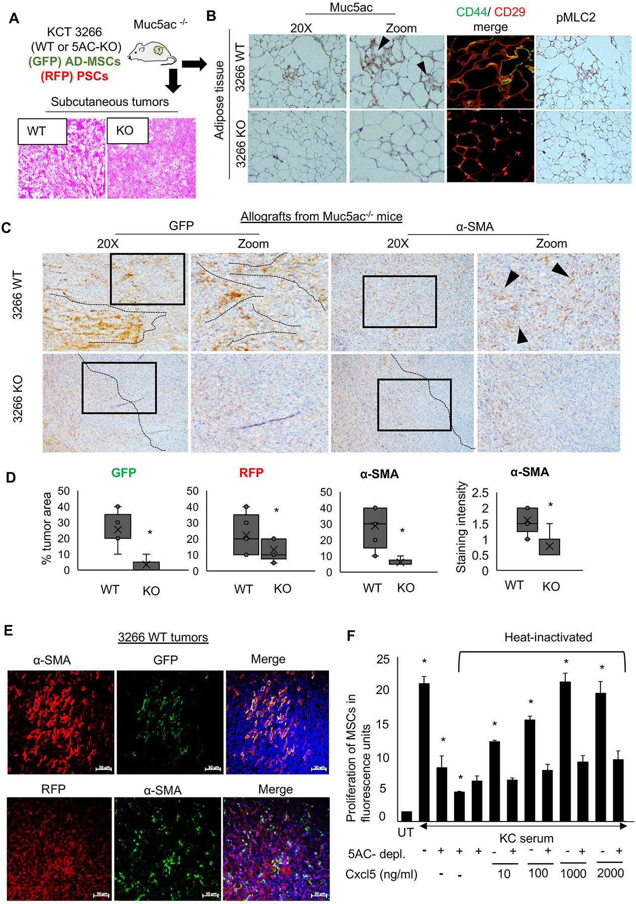
Figure 5. Muc5ac promotes AD-MSC expansion and maturation towards α-SMA-expressing CAFs.

(A) Schematic experimental plan for the co-implantation allograft model in Muc5ac−/− mice, using murine PC cell line KCT 3266 (WT and MUC5AC-KO) admixed with AD-MSCs isolated from GFP mice and RFP-labelled pancreatic stellate cells. Representative H&E images demonstrating the histology of WT and KO tumors (n=10/group). (B) Immunohistochemistry and immunofluorescence images from adipose tissue isolated from Muc5ac−/− animals demonstrate the presence of Muc5ac, with higher CD44/CD29 colocalization and pMLC2 expression in the mice grafted with Muc5ac-expressing cells, as compared to those receiving Muc5ac-KO cells. (C) Representative images and (D) quantitative analysis (n=10/group) from immunohistochemistry demonstrate the expansion of AD-MSCs (GFP), pancreatic stellate cells (RFP), and α-SMA+ CAFs with increased α-SMA staining in 3266 WT tumors, as compared to the KO group. Black dotted lines and arrowheads demonstrate the pattern of GFP-MSCs and α-SMA-CAFs, localized near the malignant cells in the WT tumors but dispersed towards the periphery of the KO tumors. (E) Immunofluorescence images show co-expression of GFP and α-SMA in the WT tumors, with no significant co-expression of RFP and α-SMA, suggesting AD-MSCs contribute more than the resident stellate cells towards the α-SMA+ stromal population in the Muc5ac-expressing pancreatic tumors. (F) The bar graph demonstrates that the proliferation of AD-MSCs significantly increased when treated with KC serum, which subsequently declined upon Muc5ac depletion and heat inactivation of KC serum. Ectopic addition of Cxcl5 at increasing concentrations of 10, 100, 1000, and 2000 ng/ml in heat-inactivated KC serum rescued the proliferation of AD-MSCs in a dose-dependent manner. t-test *p<0.05
