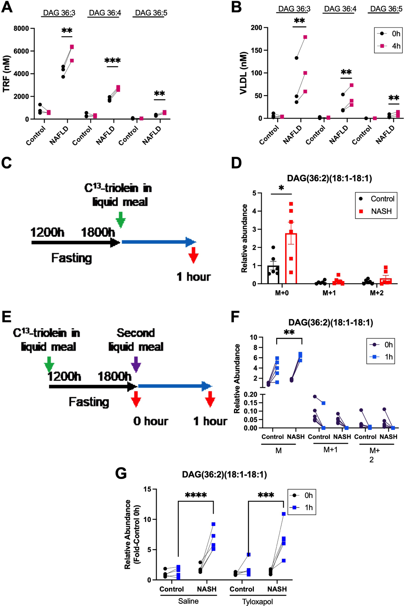
Figure 4. The postprandial increase in NAFLD plasma diacylglycerols is comprised of endogenous lipids secreted in VLDL.

(A) Plasma diacylglycerols in the triacylglycerol rich fraction (TRF) and (B) VLDL fraction at 0 and 4 hours following food bolus ingestion by control and NAFLD human subjects. (C) Schematic of 13C-triolein given in the current or (E) previous liquid meal to control and NASH mice. (D) Stable isotope labelling of postprandial plasma DAG(36:2)(18:1–18:1) in control and NASH mice 1 hour after a liquid meal bolus containing 13C-triolein. (F) Labelling of plasma DAG(36:2)(18:1–18:1) 1 hour after a second liquid meal when the prior liquid meal contained 13C-triolein. (G) Postprandial plasma DAG(36:2)(18:1–18:1) in control and NASH mice treated with saline or tyloxapol 30 minutes prior to liquid meal. M denotes unlabeled DAG; M+1, DAG with one labeled oleic acid; and M+2, DAG with both oleic acids labeled. Data are presented mean±SEM or individual values, statistical analysis was performed using a paired two-tailed Student’s t-test, n=3 (A, B), n=6 (D), n=5 (F, G) ** P<.01, *** P<.001, **** P<.0001.
