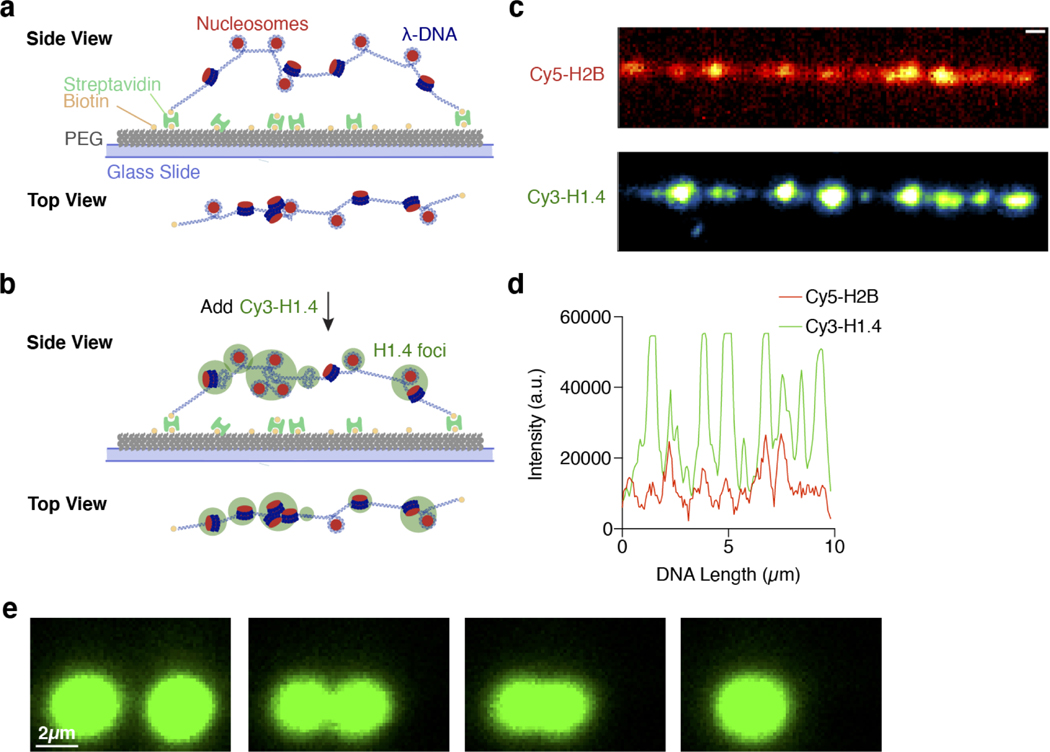
Extended Data Fig. 4. H1 colocalizes and forms condensates with nucleosomes.
a, Schematic of the total-internal-reflection fluorescence (TIRF) microscopy assay using surface-immobilized λ DNA loaded with Cy5-H2B nucleosomes. b, Schematic of the experimental setup in a after Cy3-H1 is added to the flow chamber. c, Representative fluorescence images (among 3 independentexperiments) of Cy5-H2B nucleosomes (top) and Cy3-H1 (bottom) on λ DNA. Scale bar: 0.5 μm. d, Fluorescence intensity profiles of Cy5-H2B and Cy3-H1 over the DNA length for the images in c. e, Snapshots of a representative fusion event for H1:Cy3-H2A mononucleosome droplets visualized by Cy3 fluorescence (among 21 independent fusion events).

