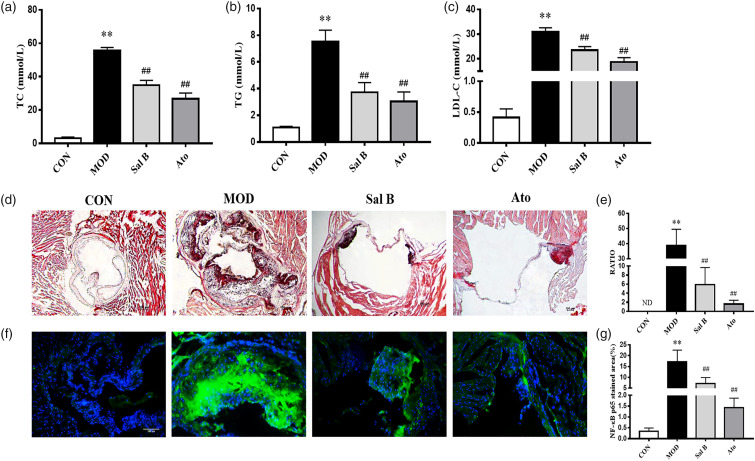
Figure 2.
Effect of Sal B on serum lipid profiles and atherosclerotic lesion area in aortic sinus of mice. (a)–(c) TC, TG, and LDL levels of serum were detected, n = 8. (d) Oil Red O staining was used to detect the progress of lipid-rich plaques, magnification ×40. (e) The areas of lesion were calculated, respectively, by ImageJ analysis software, n = 8. RATIO = plaque area/aortic sinus lumen area ×100%. (f) Immunofluorescence staining of NF-κB p65 at the aortic roots, magnification ×200. (g) The areas of NF-κB p65 stained lesion were calculated, respectively, by ImageJ analysis software, n = 3. CON, control group; MOD, model group; Sal B, Salvianolic acid B group; Ato, Atorvastatin group. TC, total cholesterol; TG, triglyceride; LDL-C, low density lipoprotein cholesterol; ND, not detected. Data are expressed as mean ± SD. **p < 0.01 vs CON group; ##p < 0.01 vs MOD group.

