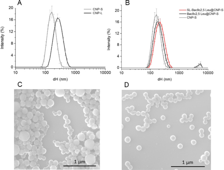
Figure 1.
Size distribution of CNPs monitored by dynamic light scattering (DLS) and scanning electron microscopy (SEM). (A) Hydrodynamic diameter distribution of CNP-S and CNP-L in water. (B) Hydrodynamic diameter distribution changes following functionalization of CNP-S with Bac8c2,5Leu or SL-Bac8c2,5Leu. Hydrodynamic diameters (dH) distribution (% intensity) is expressed as the mean value of 3 measurements ± SD. Representative SEM images of (C) CNP-L and (D) CNP-S.

