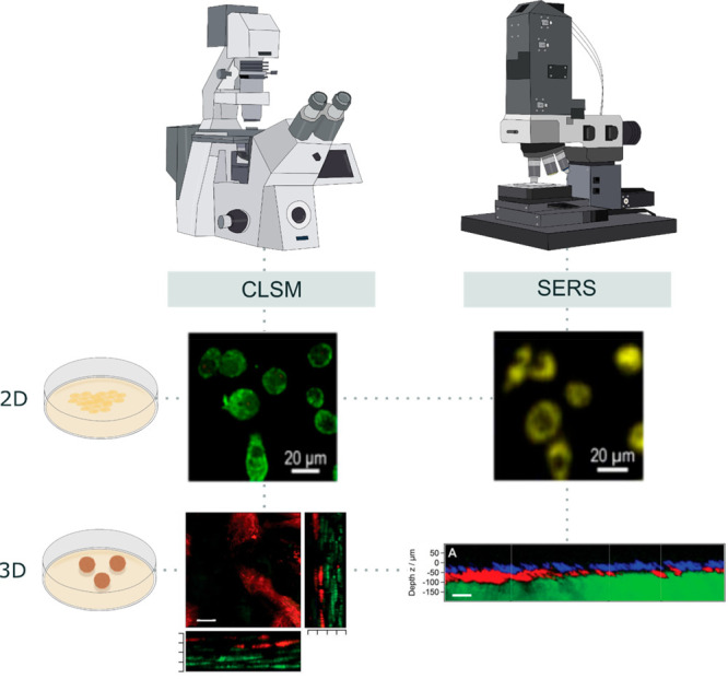
Figure 4.

Cell imaging by confocal laser scanning microscopy vs surface-enhanced Raman spectroscopy. CLSM requires from fluorescent labeling of the samples and has been traditionally used to image 3D cell cultures or tissues, but it can also image organelles at single-cell resolution. Besides the detection of various molecules, SERS can also serve as an imaging technique for cells labeled with SERS nanotags. Both 2D and 3D live-cell cultures can be imaged, guaranteeing high penetration through the sample in a less destructive way. Likewise, multiplexing is feasible with either of the aforementioned techniques, highlighting their versatility. Adapted from ref (16). Copyright 2020 American Chemical Society, and ref (17). Copyright 2020 John Wiley & Sons.
