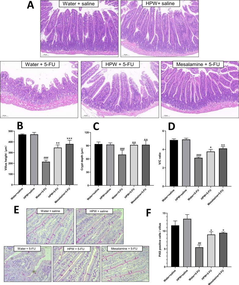
Fig. 3.
Microhistological examination of the small intestine. A HE staining of intestine tissues (scale bar = 100 μm). B Villus height. C Crypt depth. D The ratio of V/C. E PAS staining of goblet cells (scale bar = 50 μm). F Analysis of goblet cell counts. The data are presented as the means ± SEM and were analysed using one-way ANOVA followed by Dunnett’s test (n = 8). “*” represents the comparison with the model group (water + 5-FU), and “#” represents the comparison with the control group (water + saline). One tag means p < 0.05, two tokens represent p < 0.01, and three is p < 0.001

