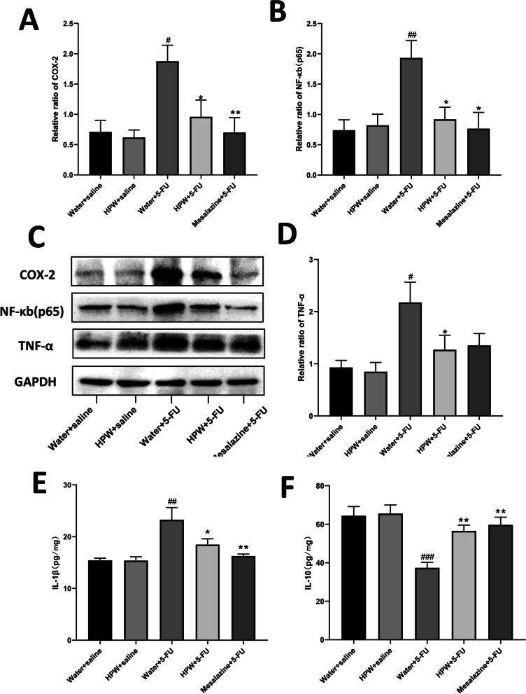
Fig. 5.
Effect of HPW on the inflammatory pathway and inflammatory factors. Greyscale value analysis of A COX-2, B NF-κB and D TNF-α. C Contrast diagram showing these three molecules and GAPDH protein bands. Analysis of E IL-1β, F IL-10 in tissue homogenate. The data are presented as the means ± SEM and were analysed using one-way ANOVA followed by Dunnett’s test (n = 8). “*” represents the comparison with the model group (water + 5-FU), and “#” represents the comparison with the control group (water + saline). One tag means p < 0.05, two tokens represent p < 0.01, and three are p < 0.001

