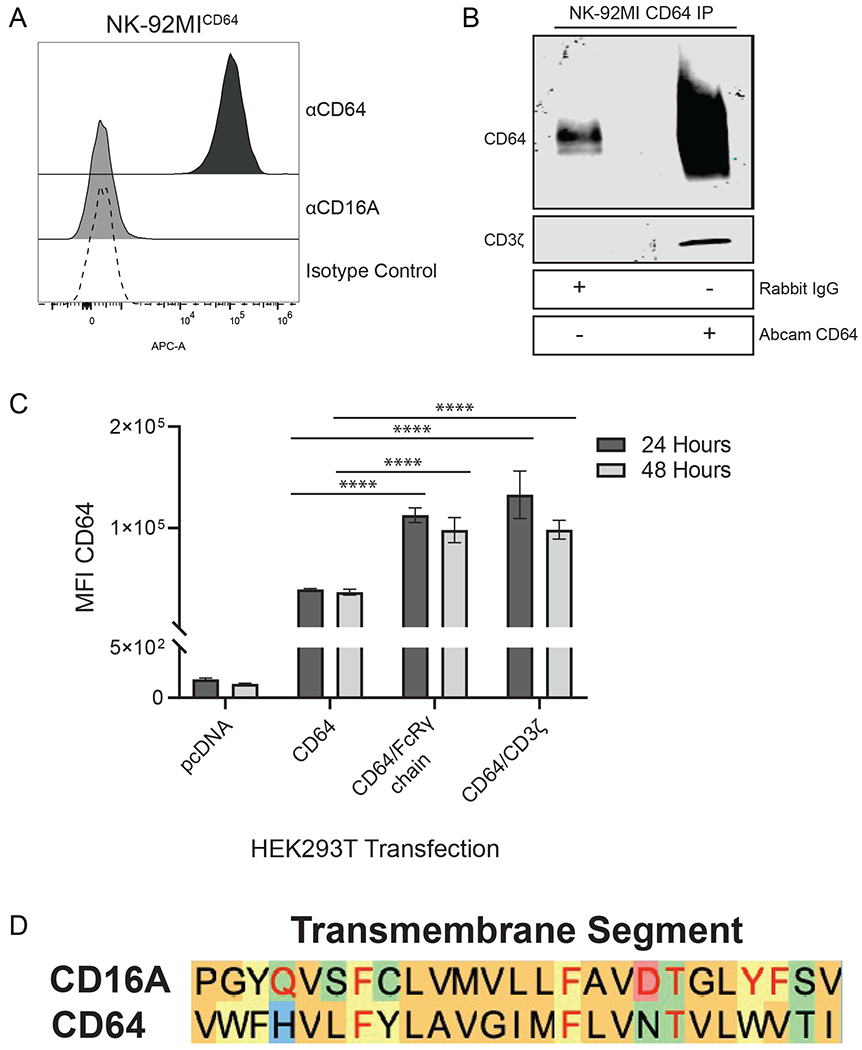
Figure 1. CD64 in transduced NK-92MI cells associates with the signaling adaptor CD3ζ.

A. CD64-transduced NK-92MI cells were stained with anti-CD64, anti-CD16, or isotype matched control antibodies and analyzed via flow cytometry. Histogram is a representative of data from 3 independent experiments. B. Lysates from NK-92MICD64 cells were immunoprecipitated with either a rabbit isotype matched control antibody or anti-CD64 and immunoblotted for CD64 and CD3ζ. Blot is a representative of 3 independent experiments. C. HEK293T cells were transiently transfected with pcDNA3.1 vector, CD64 expression construct, CD64 plus FcRγ expression constructs, or CD64 plus CD3ζ expression constructs. Cell surface expression of CD64 was determined by flow cytometry. Data are represented as mean ± SEM; n = 3 independent experiments; ****, P ≤ 0.0001 by ANOVA and Tukey’s post hoc for multiple comparisons. D. Amino acid alignment of the predicted human CD16A and human CD64 transmembrane regions. The amino acid sequences of the Fc receptor transmembrane regions are based on a previous study [50]. The color of the shaded letters represents groupings based on side chain properties. The red letters in the CD16A sequence represent amino acids critical for CD3ζ association [51]. The red letters in the CD64 sequence represent identical amino acids.
