
Fig. 1.
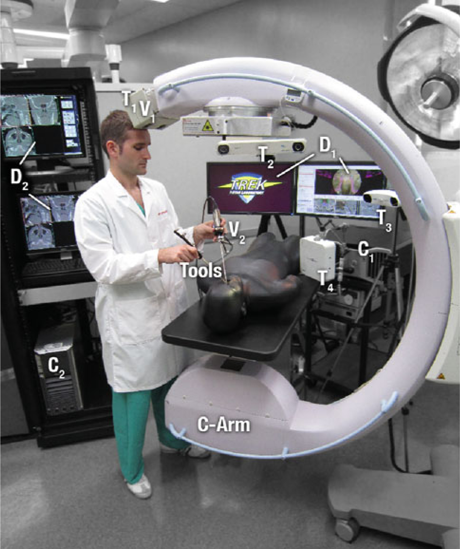
Prototype C-arm for CBCT, tracking and navigation technologies. Systems labeled in the photograph include the C-arm, tracked tools, T1 (MicronTracker, Claron, Toronto ON), T2 (Polaris Spectra, NDI, Waterloo ON), T3 (Polaris Vicra, NDI), T4 (Aurora, NDI), V1 (MicronTracker, Claron), V2 (Video endoscope, 7230AA, Karl Storz, Tuttlingen Germany), C1 (TREK navigation workstation), C2 (C-arm image acquisition workstation), D1 (TREK navigation displays), and D2 (C-arm image displays). C2/D2 are traditionally separate from the tableside setup, operated by a technologist to acquire and process C-arm images and optionally exposed to the surgeon for image review. Instead, images from C2/D2 are acquired by the technologist and transferred to C1/D1 for real-time guidance and visualization by the surgeon