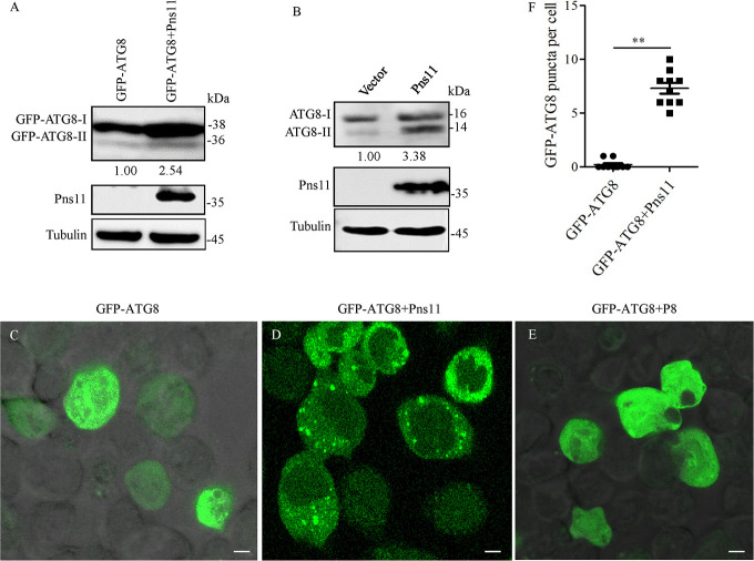
Fig 3. RGDV Pns11 induces autophagy in Sf9 cells.
(A) Accumulation levels of ATG8 and RGDV Pns11 in Sf9 cells singly expressed with GFP-ATG8 or co-expressed with GFP-ATG8 and Pns11, as detected by western blot assay. Tubulin was detected as a control. (B) Accumulation levels of ATG8 and RGDV Pns11 in Sf9 cells infected with empty or recombinant bacmids expressing Pns11, as detected by western blot assay. Tubulin was detected as a control. Relative protein levels were determined using ImageJ and the accumulation levels of ATG8-II in the controls were normalized to 1. (C-E) Immunofluorescence assay showing GFP-ATG8 (green) in Sf9 cells singly expressed with GFP-ATG8, co-expressed with GFP-ATG8 and Pns11, or co-expressed with GFP-ATG8 and P8. (F) The average number of discrete puncta of GFP-ATG8 in Sf9 cells. Bars represent means ±SD from more than 10 individual cells. Significance (**) was determined at P < 0.01. Bars, 5 μm.

