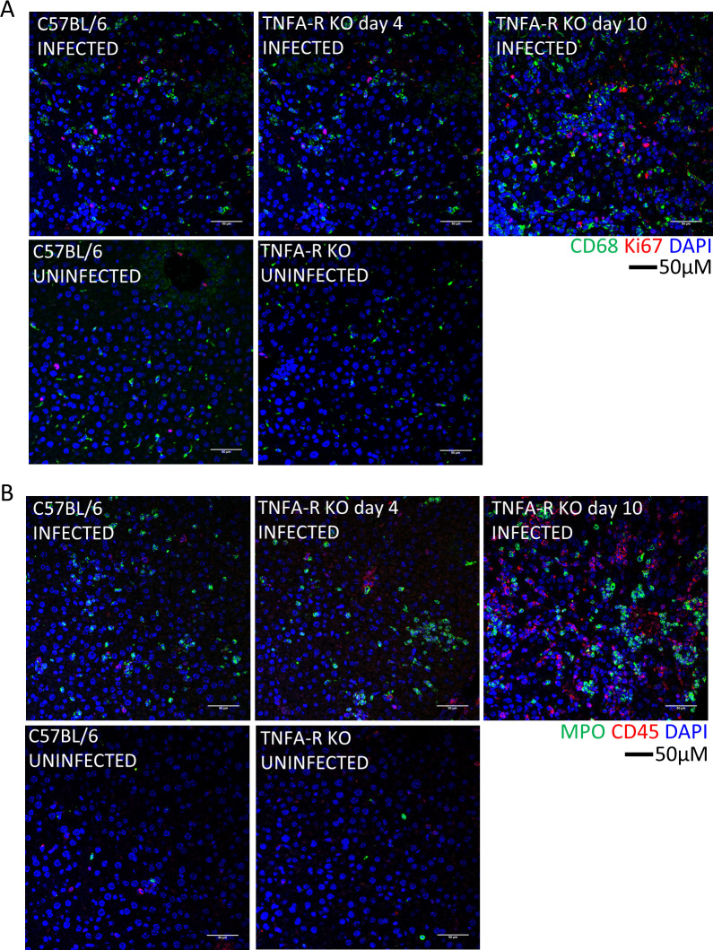
Fig 6. Immune cells in the livers of WT and TNFA-R KO mice.
Representative IFA demonstrates presence of CD68+ macrophages (A, green) and Ki67+ proliferating cells (A, red) or MPO (B, green), and CD45+ leukocytes (B, red) in livers of infected animals compared to uninfected at the indicated time points. Nuclei are stained with DAPI (blue).

