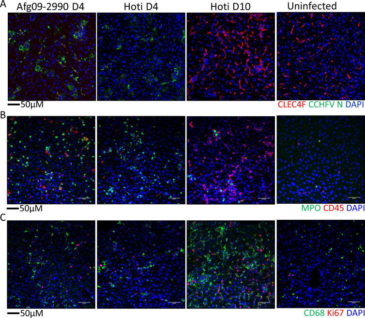
Fig 9. Immune cell levels in Hoti and Afg09-2990 infected mice.
A. Liver sections from day 4 Afg09-2990 or Hoti infected or uninfected mice were stained with anti-CLEC4F (red) and anti-CCHFV N protein (green) antibodies. B. Liver sections from were stained with MPO (green) and CD45 (red). C. IFA demonstrating presence of CD68+ macrophages (green) and Ki67+ proliferating cells (red) in livers of day 4 animals. For all panels, cell nuclei were stained with DAPI.

